
Could a diamond sensor map how electricity moves through tissue?
A $3.2 million research project aims to turn faint magnetic signals into a map of tissue conductivity. Here is what a convincing first image would show.
Reading tools for text size, copying, printing and your reading list



An imaging instrument that never touches the sample still needs a way to find out what is inside. Physicist Carlos Meriles and colleagues in New York want to try it with a diamond. Their idea is to pick up the faint magnetic response of tissue and use it to estimate where electrical current flows more easily.[1][2]
City College of New York described the collaboration on November 3. The funding predates that announcement. The federal grants ledger records a $3.2 million award on September 18, and the Advanced Research Projects Agency for Health lists the project as qSENSE. Its goal is to test whether the measurement could work well enough to build a portable imaging instrument.[1][2][3]
What would the diamond actually measure?
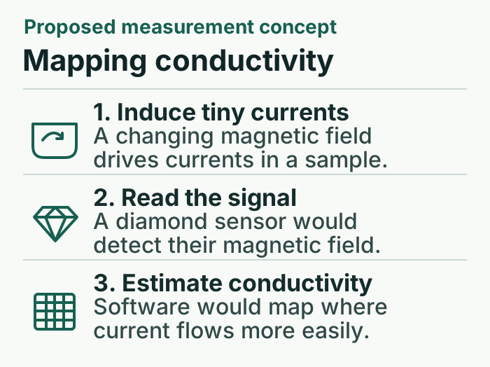
A changing magnetic field can set small currents circulating in conductive material. Those currents produce a second magnetic field that a sensitive instrument can pick up from outside. Imagine sliding a container of salt water past such an instrument. As the container moves, the field the instrument detects changes, even though the liquid never touches the sensor.[4]
Meriles’s team plans to use tiny defects in diamond called nitrogen-vacancy centers. Their response to a magnetic field can be read with light at room temperature. Qiushi Guo’s group brings expertise in building compact optical components. Andrea Alù will work on how to arrange the measurements and extract the information about position that an image requires.[1][2]
The distinction matters if you picture two samples with the same total amount of salt water. One might hold the liquid in a single pocket and the other in two pockets farther apart. Detecting a signal from either sample would be encouraging. An image has a harder job. It should put each pocket in the right place. The software must work backward from the measured fields to estimate how conductivity varies across the sample.[1][2][4]
What a useful first picture could show
There is precedent for the simpler measurement. In a separate 2024 experiment, researchers used a sensor made with alkali atoms to detect small saltwater containers at conductivities as low as 0.55 siemens per meter. That experiment gives a concrete example of the kind of weakly conducting material an instrument can detect. It used a different sensor and did not test the New York design.[5]
For qSENSE, an informative test would be a sample whose internal layout is known to the person checking the result. Can the reconstruction distinguish two nearby pockets? Does it put them at the right depth? How long does that image take to collect? Answers would tell a reader much more about imaging performance than a single number for the weakest field the sensor can detect.[1][2]
ARPA-H describes bedside use as a possibility for places where larger imaging equipment is impractical. Neither the agency’s project record nor the university’s announcement presents a scan of a patient or a measure of diagnostic accuracy. For now, the result to look for is an image whose accuracy someone can check against the object that produced it.[1][2]