
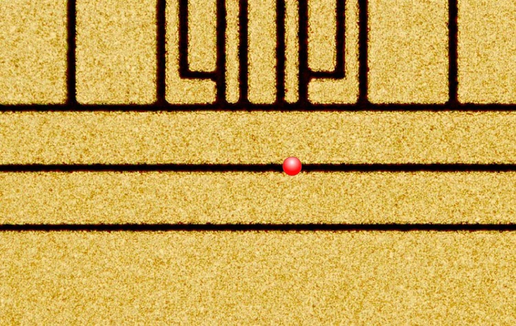
The marker is not the ion
The most important part of this caption is the status of the red dot. NIST describes it as indicating where an ion hovers above the chip. It is an annotation, not direct photographic evidence of a red atom sitting on the surface. The patterned gold electrodes are the device being imaged. This makes the file an excellent lesson in reading scientific visuals: a real micrograph can contain a teaching overlay, and each layer has a different evidential role. Preserve that distinction in alt text, credits and any crop. A companion illustration can separately show the vertical relationship between the chip and the trapped ion.
- Organization
- NIST
- Image type
- Photograph
- Capture date
- 2011-08-25
- Photographer / creator
- NIST
- Reuse terms
- Public domain
NIST — Ion Trap (6099414213).jpg — Public domain — https://commons.wikimedia.org/wiki/File:Ion_Trap_(6099414213).jpg — https://commons.wikimedia.org/wiki/File:Ion_Trap_(6099414213).jpg#Licensing.
Full original frame retained; EXIF orientation normalized where present, resized without enlargement and converted to WebP. No crop or content alteration.
Read the image
An annotation can identify a location without photographing an atom
A red circular marker sits over a patterned surface divided by dark gaps. The NIST image is an annotated micrograph: the marker explains the intended ion location. It is not a literal red atom captured at that size and brightness by the imaging system.
This distinction is central to reading scientific images. A micrograph can record physical structure, while an added symbol communicates interpretation. Both can be useful, provided a caption tells the reader which is which. Removing that distinction can make the apparatus appear to contain a directly visible object that the camera did not record.
The patterned surface is a fabrication subject; the marked location is an explanatory layer. A good gallery retains the original annotation and identifies its role. If a new explanatory diagram is needed, place it beside the source image and label it as a separate conceptual drawing.
What does the red marker mean?
It indicates the intended location of the trapped ion according to the source. Its size and color should not be interpreted as measured atomic appearance.
Why preserve the original source context?
The annotation relies on its caption. Detaching the image from that explanation can change what a reader thinks was observed.
Can a new crop remove the surrounding structure?
It may obscure why the location matters. Any crop should preserve enough context, record the modification and retain the NIST acknowledgment.
An independent QubitWire lesson based on this image and the cited source reading. The image does not establish a performance result.
Keep exploring
More from the same organization or hardware architecture.
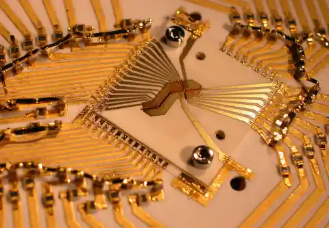 Gold electrodes for trapped ionsBlakestad/NIST · NQI gallery reuse permission
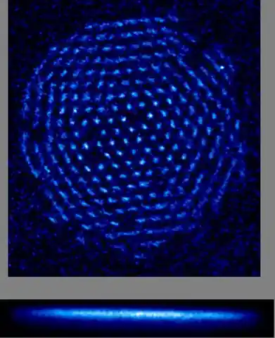 A crystal of trapped ionsNational Institute of Standards and Technology · Public domain (U.S. Government)
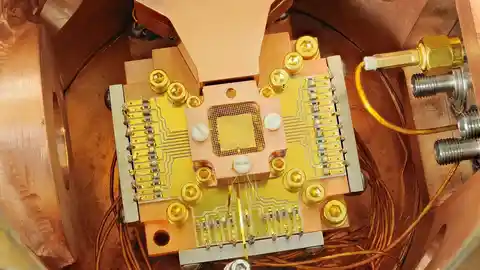 NIST’s beryllium-ion apparatusY. Colombe / NIST · Public domain (U.S. Government)
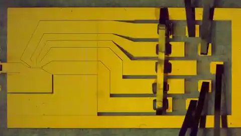 Gold electrodes for trapping ionsSigne Seidelin and John Chiaverini / NIST · Public domain (U.S. Government)
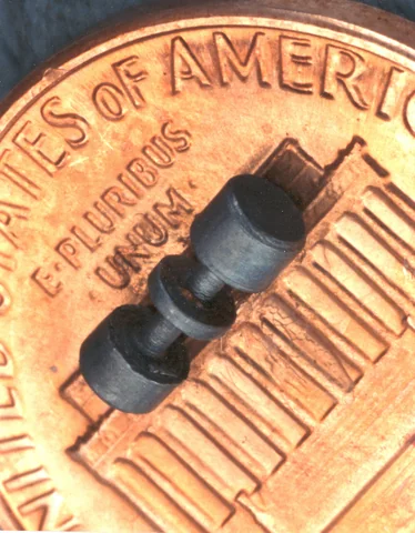 An ion trap resting on a pennyJ. Bergquist and D. Wineland / NIST · Public domain
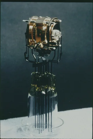 An early laser-cooling ion trapNIST · Public domain
Back to all images · Explore the interactive Model · Image rights and corrections